


















We may now know which neurons dictate when it is time to go to sleep
Two clusters of neurons in mice's brains seem to create the drive to get some shuteye, potentially hinting at a new target for sleep disorder treatments

Researchers have identified specific groups of neurons in the brains of mice that are activated after the rodents have been awake for a long time, driving them to get some sleep. If a similar system exists in humans, it could be targeted to help improve treatments for some sleep disorders.
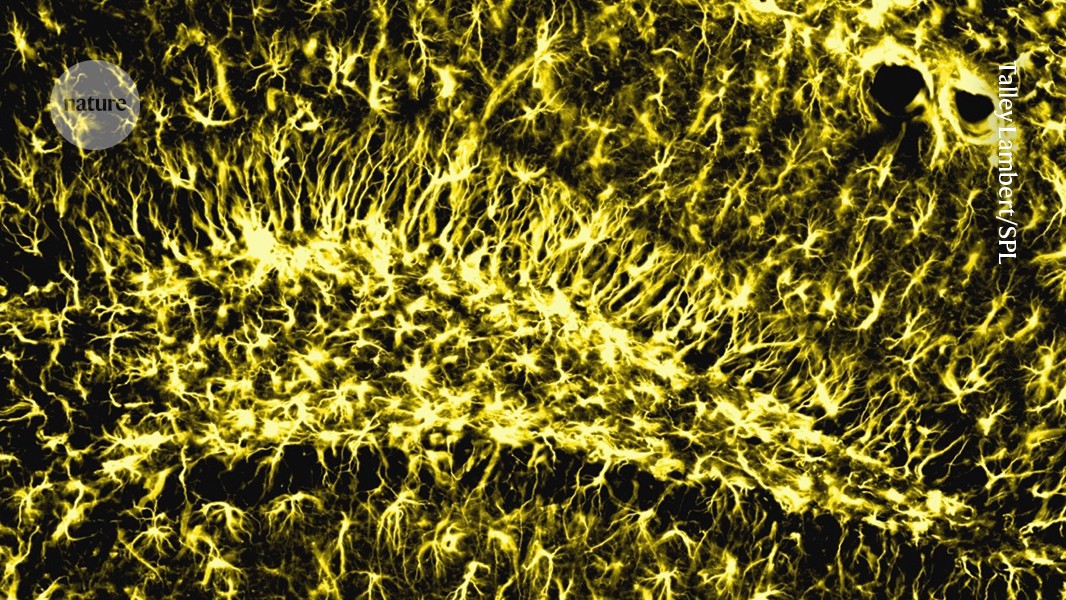
Inside the brain, several neural circuits regulate sleep and wakefulness, including neurons in the hypothalamus that act like a switchboard to control the process.
One or more of these circuits might be involved in why it is that, the longer we are awake, the more irresistible the urge to close our eyes becomes. But what actually drives this urge has long been a mystery.
Advertisement
To find out, Will Joo at the University of Basel in Switzerland and his colleagues have compared brain activation patterns in mice during normal sleep-wake cycles, sleep deprivation and in recovery sleep. They did this by tracing how much protein is produced in cells by a gene called Fos, which is turned on when neurons get activated.
“We saw that very specific parts of the brain were correlated with wakefulness,” says Joo. This allowed them to identify brain areas in which activity correlated with time spent awake.
Within one of these regions, the median raphe, they found two distinct neuronal populations that were increasingly activated the longer the animals stayed awake and which got quieter again after sleep started. These populations are the GABAergic and serotonergic neurons, so named because the chemical messenger molecules they respond to are gamma-aminobutyric acid (GABA) and serotonin, respectively.
“This is an outstanding study. The whole-brain Fos mapping is a real tour de force,” says Chiara Cirelli at the University of Wisconsin–Madison.

To see whether these neuronal populations become more active simply because the mice had been awake for a long time or whether they do something more, Joo and his colleagues next used viruses to deliver a genetic package to the neurons to either activate or inhibit their action.
When both sets of neurons were artificially activated, the mice slept for twice or three times as long as control mice and spent more time in non-REM slow-wave sleep – in a way that resembles the deeper, recovery sleep humans have after we’ve been up for a long time.
“Not only does it increase the amount of time the animal spends sleeping but it also increases the quality of the sleep,” says Joo.
And when the neurons were inhibited, the mice slept about 70 per cent less than control mice – spending about 6.5 extra hours awake a day – without showing more of the anxiety-like behaviours that usually come with sleep deprivation for mice. However, the lack of sleep was lethal in about 17 per cent of the mice.
The strong effect of activating and inhibiting the neurons means they don’t just signal that an animal has been awake, says team member Alex Schier, also at the University of Basel. “They are crucial to promote sleep, and may be key components of the neural circuitry that generates sleep drive.”
“The work adds to a growing body of work showing that there are distinct sleep circuits that are activated by wakefulness, yet promote sleep,” says Mark Wu at Johns Hopkins University in Baltimore, Maryland, whose work has shown that neurons in a brain region called the thalamic nucleus reuniens track the gap between how much sleep we need and how much we actually get.
It isn’t known if the same sleep-driving neuron populations exist in humans, but if they do, the findings could open doors to new therapies for some sleep-related conditions – once we know how the cells are communicating with other brain regions, say the researchers.
Projections from these neurons in the median raphe link to the preoptic hypothalamus, and these two areas are known to work together to control the sleep-wake cycle.
“The effect of this major neuronal path on sleep is very strong, but as we have learned since we started manipulating neuronal circuits, each path provides nuances. The brain has many ways to wake up and fall asleep,” says Luis de Lecea at Stanford University.
For example, there are hints from experiments in fruit flies that mitochondria – the powerhouses of cells – in the brain may play a crucial role in the onset of sleep.
What's Your Reaction?